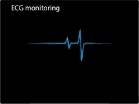
ECG monitoring
With a graphical image on the ECG we can see the electrical activity of the myocardium. Principle of ECG is based on the physical law that the movable wire that passes between the electromagnet poles turns when through her an electrical current passes. ECG is printed on millimeter paper, which makes it easy to determined the duration and amplitude of individual complexes, intervals and segments. With reading of ECG, heart rate can be determined, also rhythm, axis, and is there a hypertrophy and heart attack.
ECG monitoring is, the ECG for a period of 30-45 minutes. Often patients come with complaining of a heart skipping, or a rapid heart rate, without the prior ECG recordings that show cardiac arrhythmias. This is because patients just have done ECG and ECG tape-recording duration is only a few seconds. In such patients we need to use ECG monitoring for half an hour, because ECG takes time to show of abnormal heart rhythm. Short-term using the ECG cannot evaluated cardiac risk in people with no symptoms, such as if the patient is a smoker, if you have high blood pressure or cholesterol or a family history of heart disease. ECG is not recommended for use for patients with a low risk of heart disease.
ECG monitoring of the patient is important in the case of transient-type conduction abnormalities and ischemic heart disease.
ECG monitoring is not used to display heart deformity or for blood pressure but can display electrical activity of the heart. ECG monitoring use the electrodes that are attached to several places, usually there are 3 leads that connect to the right arm, left arm and left. leg, but there is ECG monitoring with a 12 leads.
To have a good connection between the electrodes and the skin, ECG monitoring use a gel that reduces the resistance of the skin, because it is the only way to have accurate readings. If you use the 12 leads ECG monitoring with 12 electrodes, distributed around the limbs and across the chest, you can observe many defects.
The patient upon arrival is connected with ten self-adhesive electrodes and ECG adapter that enables wireless ECG monitoring in all offices, this is followed by review and discussion with the cardiologist.
The advantage of this type of connections is the possibility of long-term monitoring.
ECG monitoring has a specific diagnostic value in following conditions:
- Disruption of the rhythm
- Disturbances in the rhythm of heart (arrhythmia or variable rhythm, tachycardia, or fast heart rate, bradycardia, or slow heart rate, and skipping heart beats, heart block, atrial chambers, atrial fibrillation)
- Hypertrophy of the heart
- Slow action, conduction of potential impulses through the atria and ventricles
- Ischemic and myocardial infarction
- Pericarditis
- Systemic diseases of the heart
- Disturbance of the balance of electrolytes
Each of these defects and disorders is reflected in a specific and distinctive way on ECG recording, and its interpretation can quickly set up a correct diagnosis.
You may also like:
- Inappropriate sinus tachycardia
Inappropriate sinus tachycardia is a heart rhythm disturbance or form of arrhythmia in which the resting heart rate is very high greater than 100 beats per minute.
- Bradycardia symptoms
Bradycardia is a heart arrhythmia that is characterized by a very slow heartbeat. Bradycardia symptoms are fatigue, weakness, shortness of breath, fainting, dizziness, memory problems and sleep problems.
- Angina pectoris – (“My heart hurts”)
Tightness or pressure as pain, retrosternal or easy to left (heart hurts). Angina pectoris occurs quickly during exertion, can extend and lose after the break.


